Quantophyse: DCE Perfusion MRI | Engineering Science Department - University of Oxford
DCE Perfusion MRI
Dynamic Contrast Enhanced Perfusion MRI
Overview
Dynamic Contrast Enhanced MRI (DCE-MRI) is an physiological MRI technique that captures the tissue T1 changes over time after the administration of a gadolinium contrast agent, revealing aspects of perfusion and permeability. DCE MRI primarily has application in providing perfusion based biomarkers in tumours.
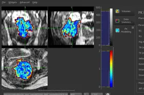
DCE MRI Analysis in Quantiphyse
Bayesian DCE Modelling
This widget provides DCE model fitting to output image maps of physiological parameters if interest such as 𝐾𝑡𝑟𝑎𝑛𝑠, 𝐹𝑝, 𝑃𝑆, 𝑉𝑝 and 𝑉𝑒. A Bayesian inference approach is used, this has the advantage that prior knowledge about likely parameter values can be incorporated. This allows more complex models to be implemented and is particularly well suited to data with high temporal sampling.
Models included:
- The standard and extended one-compartment Tofts model
- The two-compartment exchange model (2CXM)
- The Compartmental Tissue Uptake model (CTU)
- The Adiabatic Approximation to the Tissue Homogeniety model (AATH)
AIF options
- Specified from the data (using Quantiphyse tools)
- User supplied
- Population models (Orton, Parker)
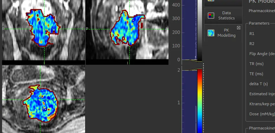
Least-squares DCE Modelling
Pharmacokinetic modelling for Dynamic Contrast-Enhanced MRI (DCE) using the Tofts model and least-squares model fitting. Less flexible than the Bayesian modelling tool, but consistent with many literature studies on conventional DCE data.
Models included:
- Tofts model
AIF options
- Orton population model (clinical)
- Heilman population model (preclinical)